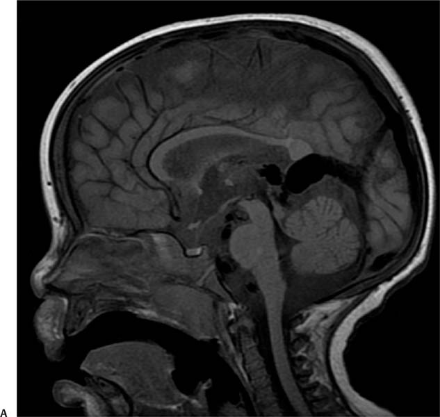
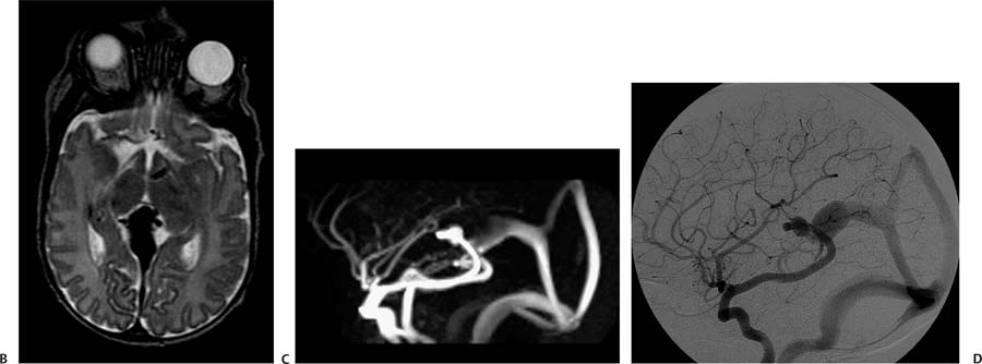
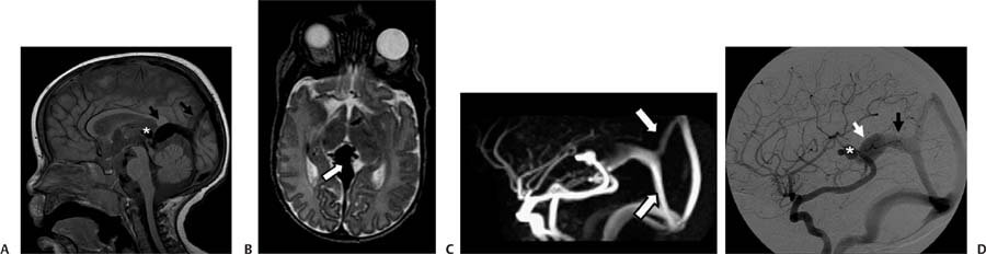
Case 67 A premature infant with cardiac failure. (A) Sagittal T1-weighted image (WI) shows large flow voids in the region of the tectum (asterisk) and prominent midline veins in the margin of the tentorium and occipital region (arrows). (B) Axial T2WI confirms prominent veins in the tectal region (arrow). (C) Sagittal phase-contrast magnetic resonance (MR) venogram shows enlarged veins from the tectum to the torcula and sagittal sinus (arrows). (D) Digital subtracted angiogram of a left internal carotid injection shows a prominent posterior communicating artery and posterior cerebral artery shunting into a venous pouch (asterisk) located anterosuperiorly to the dilated vein of Galen (white arrow), which subsequently drains into a falcine sinus (black arrow) toward the sagittal sinus and torcula. • Vein of Galen aneurysmal malformation (VGAM):
Clinical Presentation
Further Work-up
Imaging Findings
Differential Diagnosis
![]()
Stay updated, free articles. Join our Telegram channel

Full access? Get Clinical Tree


