 Clinical Presentation
A 49-year-old woman with muscle weakness.
 Imaging Findings
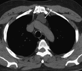
Computed tomography (CT) of the chest (soft-tissue window) demonstrates a well-circumscribed, smooth, hypodense mass in the anterior mediastinum (arrow). There is no evidence of vascular or pleural involvement.
 Differential Diagnosis
• Thymoma: This is the most common anterior mediastinal mass, especially in patients older than 40 years of age. The homogeneous density, smooth borders, and lack of local invasion support thymoma. The clinical history suggests the presence of myasthenia gravis.
• Germ cell tumor:
Stay updated, free articles. Join our Telegram channel

Full access? Get Clinical Tree


