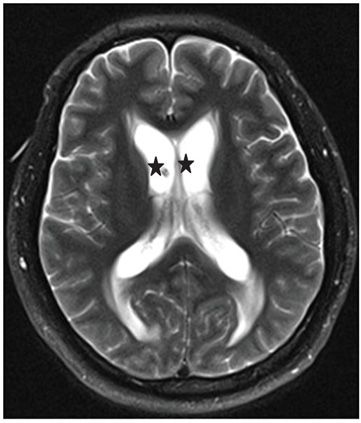
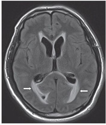
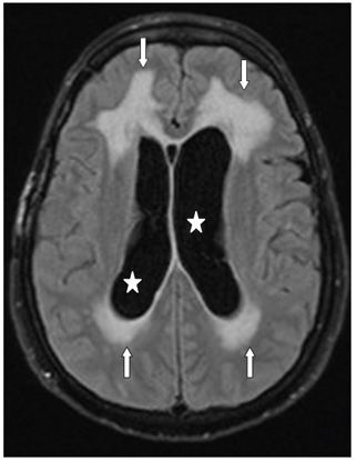
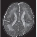
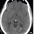
FINDINGS Figure 67-1. Axial NCCT through the lateral ventricles shows dilatation of the lateral ventricles (stars) and absence of cortical sulci. There is subtle periventricular hypodensity (arrows). Figures 67-2 to 67-4. Corresponding axial T2WI and FLAIR through various levels of the lateral ventricles show the dilated ventricles (star) and confluent periventricular hyperintensity (arrows) which is better demonstrated on the FLAIR images due to cerebrospinal fluid (CSF) suppression. This confluent periventricular hyperintensity in the setting of hydrocephalus is known as periventricular edema or transependymal CSF flow.
DIFFERENTIAL DIAGNOSIS Small-vessel ischemic disease (leukoaraiosis), cerebral volume loss, normal pressure hydrocephalus, communicating hydrocephalus, noncommunicating hydrocephalus, periventricular lymphoma, ependymitis granularis.
DIAGNOSIS Transependymal CSF flow or edema due to obstructive hydrocephalus.
DISCUSSION


Stay updated, free articles. Join our Telegram channel

Full access? Get Clinical Tree


