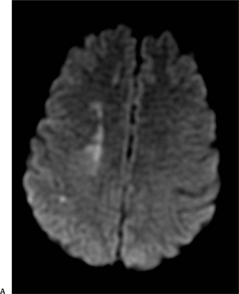
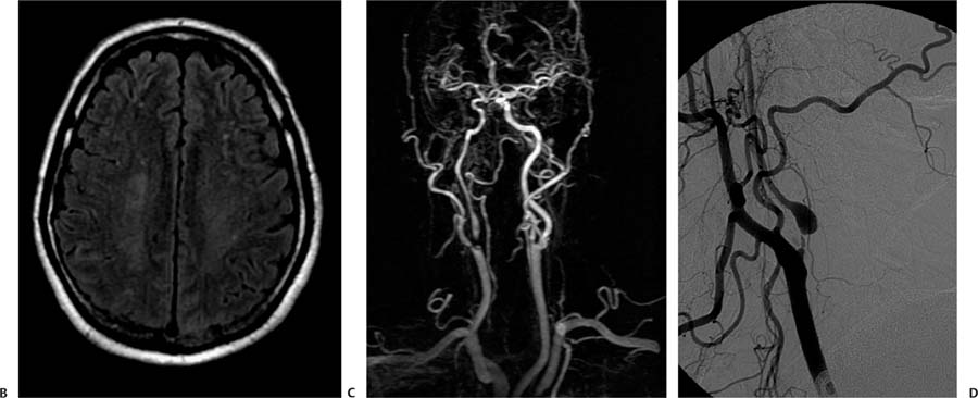
Case 68 A 75-year-old woman presenting with transient left hemiparesis. (A) diffusion image demonstrates ischemic changes in the so-called internal watershed in the deep white matter between the territories of the anterior cerebral artery and middle cerebral artery on the right (arrows). (B) Very subtle T2 signal changes are present in the same area on the fluid-attenuated inversion recovery image (arrows). (C) Magnetic resonance angiography (MRA) of the neck with contrast demonstrates signal dropout in the proximal right internal carotid artery (ICA), which is consistent with critical stenosis (arrow). The degree of stenosis cannot be quantified. (D) Critical stenosis (>90%) in the proximal ICA is confirmed on the digital subtraction angiogram (DSA; arrow). The diameter of the distal ICA (arrowheads) is small compared with that of the contralateral ICA (not shown). • Critical ICA stenosis: Infarcts in the watershed territories may result solely from hemodynamic compromise, but they are more frequent in the setting of carotid stenosis. • Carotid pseudoaneurysm:
Clinical Presentation
Further Work-up
Imaging Findings
Differential Diagnosis
![]()
Stay updated, free articles. Join our Telegram channel

Full access? Get Clinical Tree


