 Clinical Presentation
A 38-year-old woman with dyspnea.
Further Work-up

 Imaging Findings

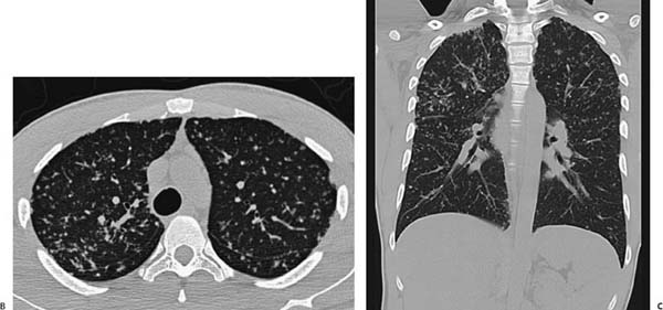
(A) Chest radiograph demonstrates upper lobe–predominant reticulonodular interstitial abnormality (arrows). There is no significant lymphadenopathy. (B) Computed tomography (CT) of the chest (lung windows) shows perilymphatic nodules and beading of the fissures. There is thickening of the bronchovascular bundles (arrows). (C) CT of the chest (coronal lung windows) shows the upper lobe distribution to advantage (arrows).
 Differential Diagnosis
• Sarcoidosis: Upper lobe–predominant nodules in a perilymphatic distribution are suggestive of sarcoidosis.
• Hypersensitivity pneumonitis (HP): HP also demonstrates relative basal sparing; however, the nodules are classically centrilobular, and more ground-glass opacity is expected. An appropriate history of exposure is sometimes present.
• Respiratory bronchiolitis:
Stay updated, free articles. Join our Telegram channel

Full access? Get Clinical Tree


