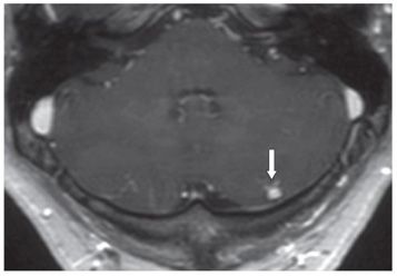
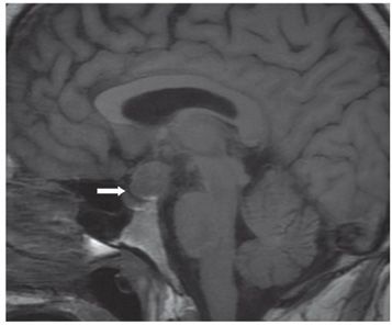
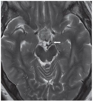
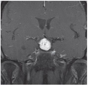
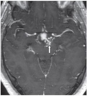
FINDINGS Figure 68-1. Axial T2-weighted MRI through the lower cerebellum. There are two small hyperintense cerebellar lesions: adjacent to the fourth ventricle on the right (transverse arrow) and posterior/peripheral on the left (vertical arrow). There is no surrounding edema. Figure 68-2. Post-contrast axial T1WI through inferior cerebellum. There is a spiculated contrast enhancement of the left cerebellar lesion (arrow). Figure 68-3. Sagittal T1WI. There is a well-circumscribed isointense suprasellar mass (arrow). Figure 68-4. Axial T2WI through the suprasellar region. The mass is heterogeneous but predominantly isointense. There are some vascular signal voids superiorly to the left of the lesion (arrow). Figure 68-5. Coronal post-contrast T1WI through the mass. The mass has avid contrast enhancement. Figure 68-6. Axial post-contrast T1WI just superiorly to the mass. There are tubular contrast-enhancing structures superolateral to the left of the lesion consistent with blood vessels (arrow).
DIFFERENTIAL DIAGNOSIS


Stay updated, free articles. Join our Telegram channel

Full access? Get Clinical Tree


