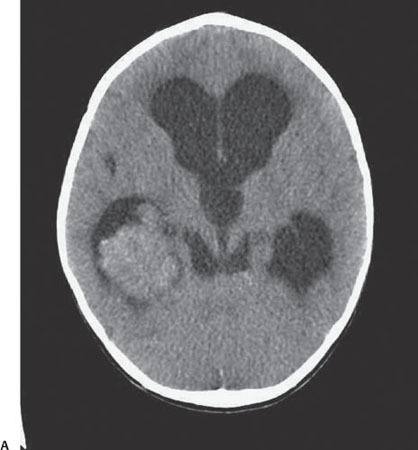
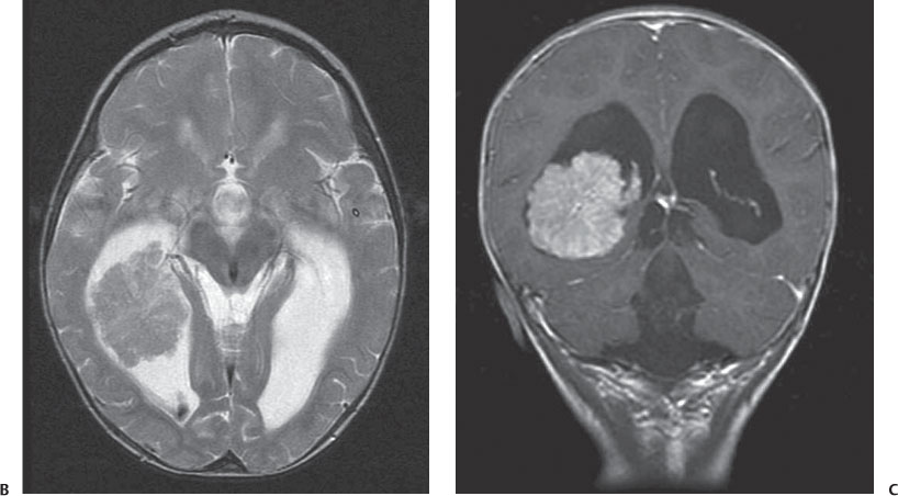
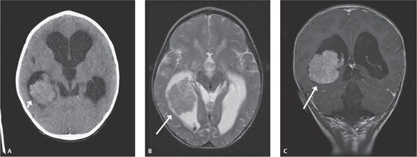
Case 68 An 8-month-old with an enlarging head circumference. (A) Nonenhanced axial computed tomography image: there is a mildly hyperdense lobular mass in the right lateral ventricle (arrow), with enlargement of the lateral and third ventricles. (B) Axial T2 magnetic resonance image (MRI): there is a well-defined, lobular, isointense mass in the lateral ventricle (arrow), with enlargement of the ventricles. (C) Coronal T1 post-contrast MRI: there is marked enhancement of the mass (arrow). • Choroid plexus papilloma (CPP)/carcinoma (CPCa): These findings are typical of CPP/CPCa.
Clinical Presentation
Further Work-up
Imaging Findings
Differential Diagnosis
Stay updated, free articles. Join our Telegram channel

Full access? Get Clinical Tree


