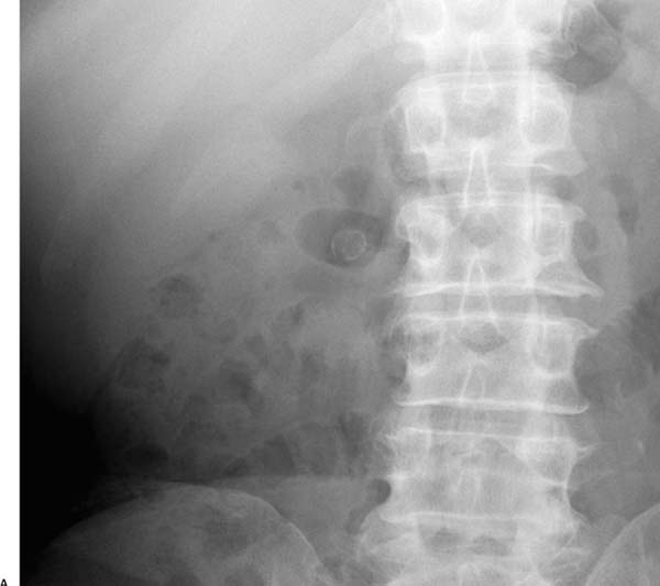
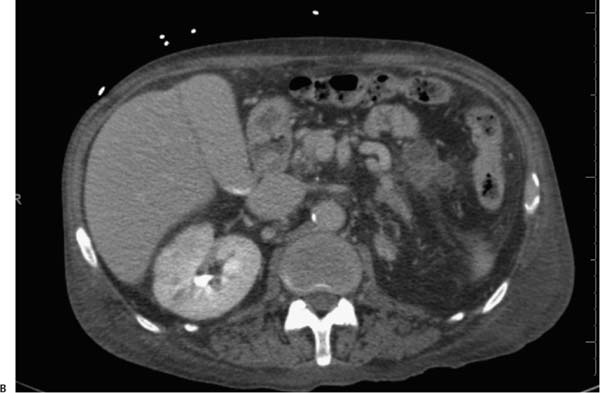
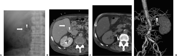
Case 68 A 68-year-old man presents for the evaluation of right upper quadrant pain. (A) Frontal abdominal radiograph shows a gas-filled structure in the right upper quadrant (arrow) superimposed on a small, round, calcified structure (arrowhead). (B) Contrast-enhanced computed tomography (CT) shows the gallbladder (arrow) to be fluid-filled (not gas-filled), with small, layering stones (arrowhead) but no large, calcified gallstones. (C) Noninfused CT shows a rounded, calcified structure adjacent to the renal artery (arrowhead). (D) CT angiography verifies renal artery aneurysm (arrowhead). • Aneurysm:
 Clinical Presentation
Further Work-up
 Imaging Findings
 Differential Diagnosis
![]()
Stay updated, free articles. Join our Telegram channel

Full access? Get Clinical Tree


