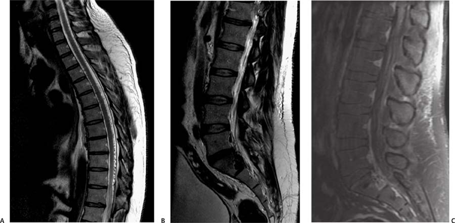
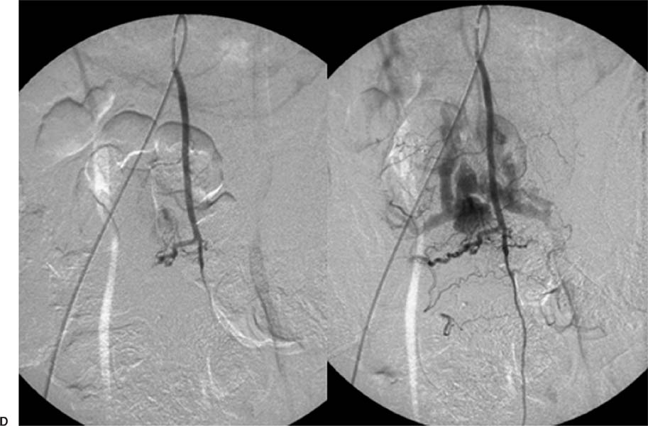
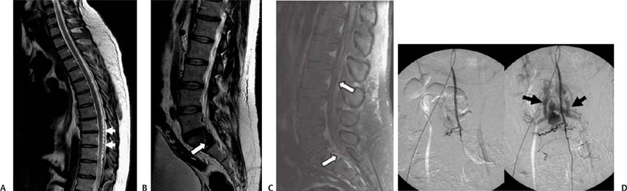
Case 69 A 38-year-old man with lower extremity numbness and progressive weakness. (A) Sagittal T2–weighted image (WI) of the thoracic spine shows multiple tiny flow voids in the posterior subarachnoid space (arrows). (B) Sagittal T2WI of the lumbar spine shows a tangle of vessels causing scalloping of the posterior margin of the S1 vertebral body (arrow). (C) Sagittal postcontrast T1WI in the lumbar region shows enhancement of the vascular structures within the thecal sac and epidural space (arrows). (D) Lateral projection of a spinal angiogram with injection in the artery of Adamkiewicz shows early opacification of large veins (arrows) in the epidural and subarachnoid spaces. No nidus is identified. • Spinal dural arteriovenous fistula:
Clinical Presentation
Further Work-up
Imaging Findings
Differential Diagnosis
![]()
Stay updated, free articles. Join our Telegram channel

Full access? Get Clinical Tree


