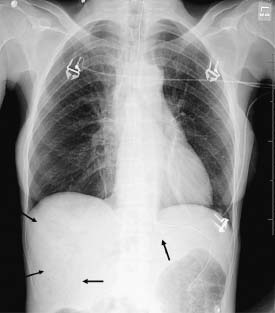
Clinical Presentation A 38-year-old woman with sepsis. Imaging Findings Chest radiograph demonstrates branching linear lucencies in the liver, dilated bowel loops, and pneumatosis intestinalis (arrows). The lungs are normal. Differential Diagnosis • Portal venous gas: Only gold members can continue reading. Log In or Register to continue Share this:Click to share on Twitter (Opens in new window)Click to share on Facebook (Opens in new window) Related posts: 15 28 50 59 86 93 Stay updated, free articles. Join our Telegram channel Join Tags: Thoracic Imaging Feb 28, 2016 | Posted by admin in RESPIRATORY IMAGING | Comments Off on 69 Full access? Get Clinical Tree