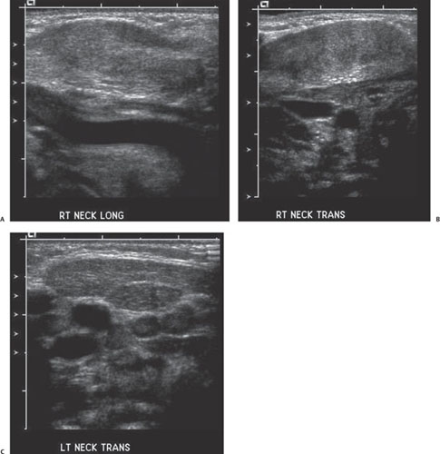
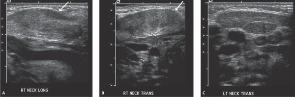
Case 69 A 2-week-old boy with a firm mass in his neck and his head turned to the left. (A,B) Longitudinal and transverse ultrasound (US) images of the right side of the neck: The right sternocleidomastoid (SCM) muscle is enlarged (arrows). There is a subtle alteration of the echotexture in comparison with the normal left side. The fascial planes are intact. (C) Transverse US image of the left side of the neck: there is a normal-appearing left SCM muscle. • Fibromatosis colli: These findings are typical of fibromatosis colli. • Cervical lymphadenopathy: This usually has more of a lobulated contour with an echogenic/fatty hilum. A conglomeration of lymph nodes should have a more distorted architecture.
Clinical Presentation
Imaging Findings
Differential Diagnosis
Stay updated, free articles. Join our Telegram channel

Full access? Get Clinical Tree


