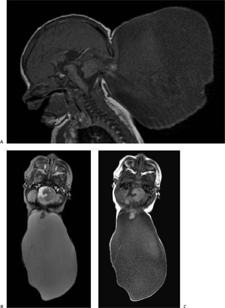
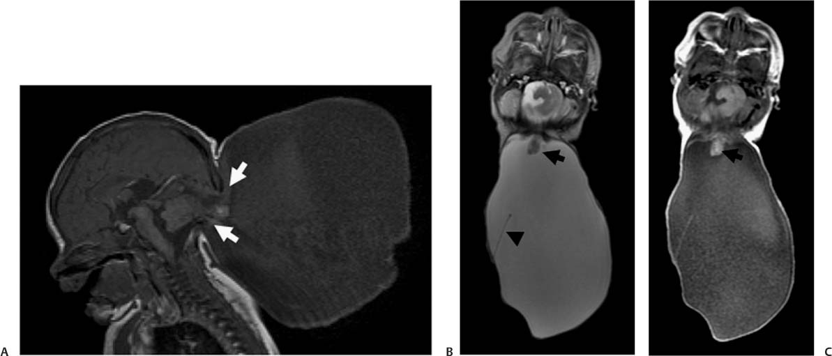
Case 7 A newborn with a posterior neck mass diagnosed on prenatal ultrasound. (A) Sagittal T1-weighted image of the brain shows a posterior skull defect (arrows) through which cerebellar tissue and cerebrospinal fluid are protruding. (B) Axial T2WI of the brain demonstrates elongation of the cerebellum, which protrudes toward the neck (arrow) and is surrounded by a large cystic lesion. Note a thin septum within the cyst (arrowhead). (C) Axial T1WI of the brain demonstrates elongation of the cerebellum, which protrudes toward the neck (arrow
Clinical Presentation
Imaging Findings
![]()
Stay updated, free articles. Join our Telegram channel

Full access? Get Clinical Tree


