 Clinical Presentation
A 26-year-old woman with progressive shortness of breath.
 Imaging Findings
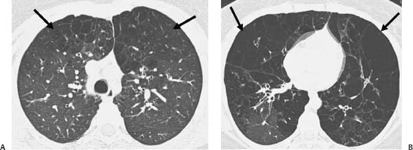
(A, B) High-resolution computed tomography of the chest: Axial images at the level of the upper lobes (A) and lower lung zones (B) demonstrate extensive emphysematous changes. Large bullous formations are seen bilaterally, more extensively involving the lower lung zones (arrows).
 Differential Diagnosis
• α1-Antitrypsin (AAT) deficiency: Panlobular emphysema, characterized by more extensive involvement of the lower lobes and lower segments in relatively young adults, is suggestive of AAT deficiency.
• Centrilobular emphysema:
Stay updated, free articles. Join our Telegram channel

Full access? Get Clinical Tree


