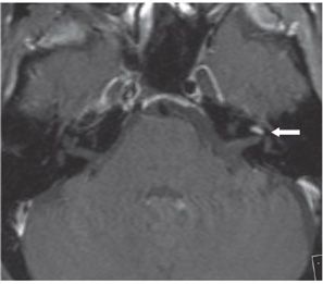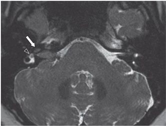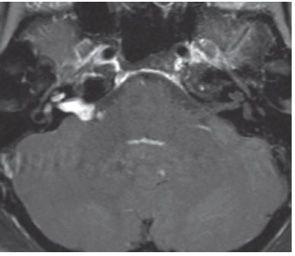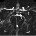


FINDINGS Figure 7-1. Axial T2WI through the cerebellopontine angles (CPAs). The normal T2 signal hyperintensity of the fluid (perilymph and endolymph) in the middle turn of the left cochlea has been replaced (arrow) (note the normal cochlea on the right, open arrowhead). Figure 7-2. Axial post-contrast T1WI through same level. There is avid corresponding enhancement (arrow). Figures 7-3 and 7-4. Axial T2WI and corresponding post-contrast T1WI through the internal auditory canal (IAC), respectively, in a companion case. There is avidly contrast-enhancing isointense tumor filling the right IAC and protruding into the CPA cistern with extension into the cochlea (transmodiolar type—arrow in Figure 7-3) and vestibule (transmacular type—arrowhead in Figure 7-3).
DIFFERENTIAL DIAGNOSIS Cochleitis/labyrinthitis, cochlear schwannoma.
DIAGNOSIS Intralabyrinthine (cochlear) schwannoma (ILS).
DISCUSSION
Stay updated, free articles. Join our Telegram channel

Full access? Get Clinical Tree


