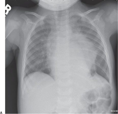
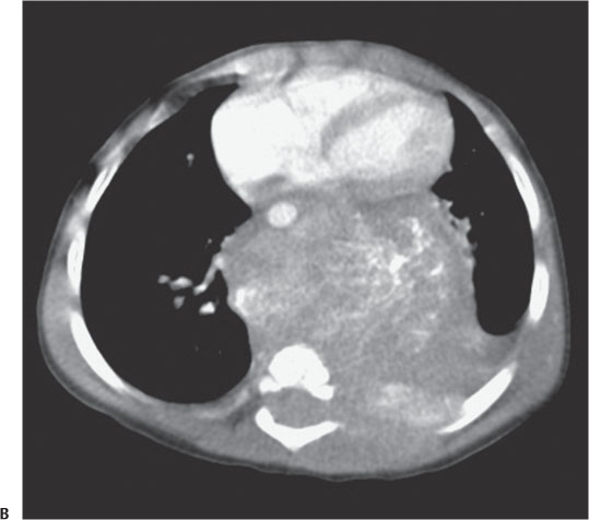
Case 7 A 3-year-old child with back pain and shortness of breath. (A) Frontal chest radiograph demonstrates a large, predominately left-sided posterior mediastinal mass. The posterior location of the mass is apparent from several features: It obscures the margin of the descending aorta, and it is associated with increased distances between the posterior aspects of the lower ribs on the left in comparison with those on the right. Moreover, these ribs are eroded and narrowed medially (arrows). (B) Axial post-contrast computed tomography image demonstrates a large posterior mediastinal mass displacing the mediastinal structures anteriorly. It contains multiple calcifications and expands the left neural foramen as it invades the vertebral canal (arrow).
Clinical Presentation
Further Work-up
Imaging Findings
Stay updated, free articles. Join our Telegram channel

Full access? Get Clinical Tree


