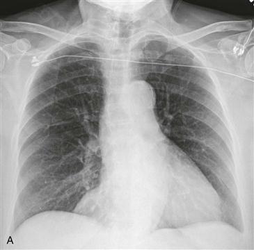
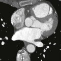
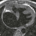
CASE 7 History: A patient undergoes preoperative chest radiography before cardiac surgery. 1. What should be included in the differential diagnosis for Fig. A? (Choose all that apply.) A. Fat pad B. Bronchogenic cyst C. Pericardial cyst D. Esophageal duplication cyst E. Bochdalek hernia 2. Where does this lesion most commonly occur? A. Right cardiophrenic angle B. Subcarinal space C. Anterior mediastinum Only gold members can continue reading. Log In or Register to continue Share this: Share on X (Opens in new window) X Share on Facebook (Opens in new window) Facebook Related posts: 11 25 51 68 90 145 Stay updated, free articles. Join our Telegram channel Join Tags: Cardiac Imaging Case Review Series Dec 26, 2015 | Posted by admin in CARDIOVASCULAR IMAGING | Comments Off on 7 Full access? Get Clinical Tree
CASE 7 History: A patient undergoes preoperative chest radiography before cardiac surgery. 1. What should be included in the differential diagnosis for Fig. A? (Choose all that apply.) A. Fat pad B. Bronchogenic cyst C. Pericardial cyst D. Esophageal duplication cyst E. Bochdalek hernia 2. Where does this lesion most commonly occur? A. Right cardiophrenic angle B. Subcarinal space C. Anterior mediastinum Only gold members can continue reading. Log In or Register to continue Share this: Share on X (Opens in new window) X Share on Facebook (Opens in new window) Facebook Related posts: 11 25 51 68 90 145 Stay updated, free articles. Join our Telegram channel Join Tags: Cardiac Imaging Case Review Series Dec 26, 2015 | Posted by admin in CARDIOVASCULAR IMAGING | Comments Off on 7 Full access? Get Clinical Tree