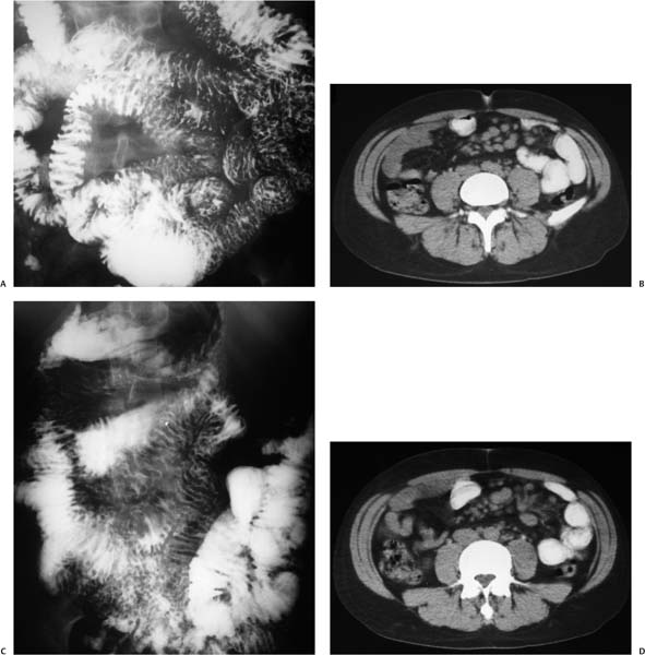
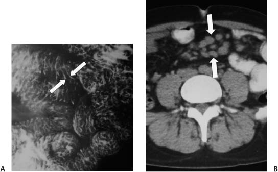
Case 7 A 52-year-old man presents with severe diarrhea. (A) Small-bowel (SB) follow-through study shows diffusely thickened, irregular, nodular jejunal folds (between arrows). (B) Axial computed tomography (CT) after oral contrast shows a cluster of enlarged mesenteric lymph nodes in the midabdomen (arrows). • Mycobacterium avium-intracellulare (MAI) infection: Irregular, nodular fold thickening, adenopathy, and diarrhea all point to MAI infection as the correct diagnosis. • Whipple disease: Whipple disease can present with similar imaging and clinical findings, but with fatty, bulkier adenopathy. • Lymphoma: Lymphoma can cause diffuse fold thickening, but typically the adenopathy will be more prominent.
 Clinical Presentation
 Imaging Findings
 Differential Diagnosis
 Essential Facts
Stay updated, free articles. Join our Telegram channel

Full access? Get Clinical Tree


