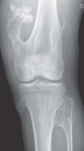
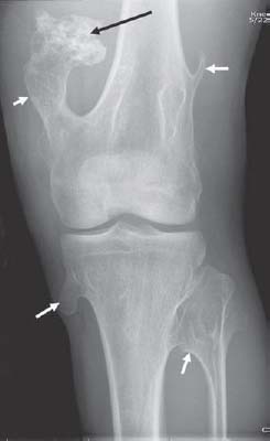
Case 70 A patient presents with a lower extremity deformity and a family history of abnormal bones. Radiograph of the left knee show multiple bony protuberances (white arrows) extending from the distal femur and proximal tibia and fibula. The lesions are continuous with the cortical and medullary bone and point away from the knee joint. No soft-tissue mass is seen. Calcifications over the femoral lesion (black arrow) are seen.

 Clinical Presentation
 Imaging Findings

Stay updated, free articles. Join our Telegram channel

Full access? Get Clinical Tree


