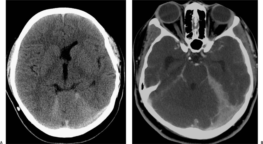
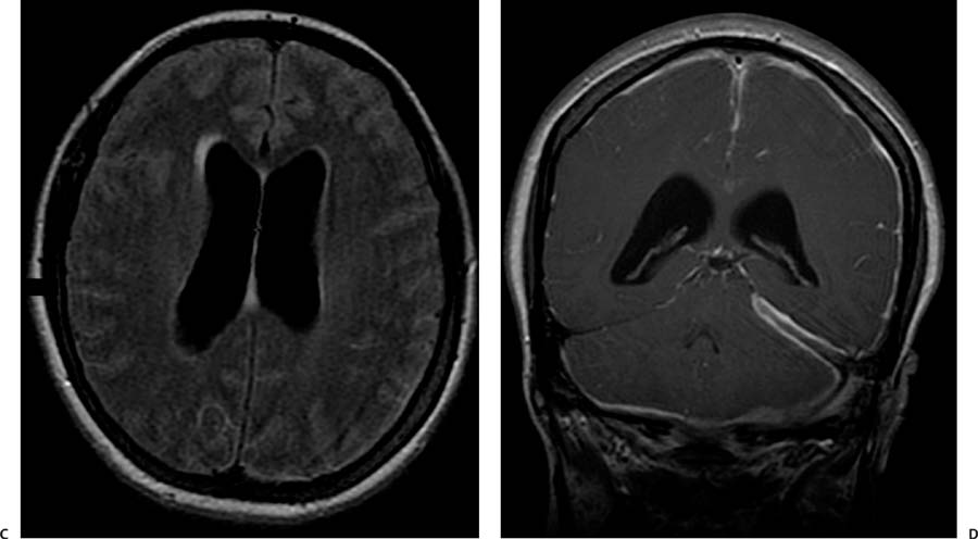
Case 70 A 17-year-old boy with progression of severe headache and lethargy that started 2 days prior. (A) Computed tomography (CT) without contrast shows effacement of the sulci (white arrows) and hyperdensity along the tentorium (black arrow). (B) CT with contrast shows diffuse meningeal enhancement that is more prominent on the left tentorium (black arrow). (C) Axial fluid-attenuated inversion recovery (FLAIR) image shows dilated ventricles with increased signal in the leptomeninges (arrows). (D) Coronal T1-weighted image (WI) with contrast shows diffuse enhancement of the meninges, more prominent on the left side (arrows
Clinical Presentation
Further Work-up
Imaging Findings
![]()
Stay updated, free articles. Join our Telegram channel

Full access? Get Clinical Tree


