 Clinical Presentation
A 52-year-old man with chest pain and hypotension.
Further Work-up

 Imaging Findings

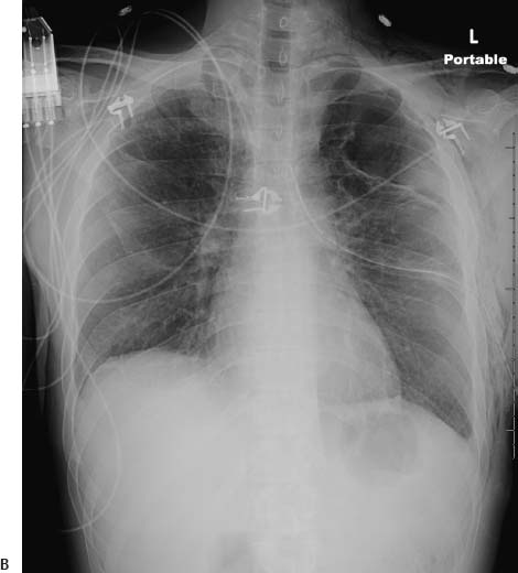
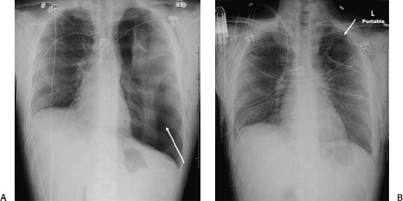
(A) Portable chest radiograph demonstrates hyperlucency of the left hemithorax with rightward mediastinal shift (arrow). There is flattening of the left diaphragm and increased space between the ribs. The left pleural line is well seen, although there are multiple pleural adhesions. Subcutaneous emphysema is present on the left. (B) Following placement of a chest tube, the left lung has expanded and the mediastinal shift has resolved. There is minimal residual pneumothorax, as evidenced by the pleural line seen at the apex of the left lung (arrow). Note the underlying emphysema and bullous changes.
 Differential Diagnosis
• Tension pneumothorax:
Stay updated, free articles. Join our Telegram channel

Full access? Get Clinical Tree


