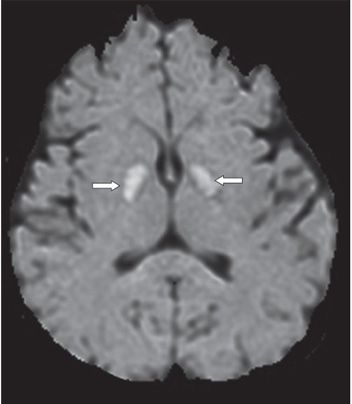
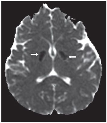
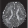
FINDINGS Figure 70-1. Axial MR T2WI through the basal ganglia. There is bilateral symmetrical globus pallidus hyperintensity (arrows). Figures 70-2 and 70-3. Axial DWI with corresponding ADC map through the basal ganglia. There is bilateral symmetrical restricted diffusion in the globus pallidus (arrows).
DIFFERENTIAL DIAGNOSIS Wilson disease, Creutzfeld-Jakob disease, Japanese encephalitis, Carbon monoxide (CO) poisoning and Leigh syndrome and other mitochondriopathies.
DIAGNOSIS Carbon monoxide (CO) poisoning.
DISCUSSION


Stay updated, free articles. Join our Telegram channel

Full access? Get Clinical Tree


