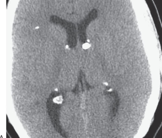
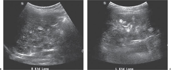
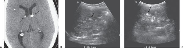
Case 70 A 10-year-old boy with seizures. (A) Axial noncontrast computed tomography (CT) image: there are multiple calcified subependymal nodules (SENs, arrows). (B,C) Ultrasound images of the kidneys: there are multiple small hyperechoic nodules scattered throughout both kidneys (arrows). • Tuberous sclerosis: Based on the calcified SENs and the echogenic lesions (angiomyolipomas) in the kidneys, this is the best diagnosis. • Sequela of a TORCH (toxoplasmosis, other infections, rubella, cytomegalovirus infection, herpes simplex) infection:
Clinical Presentation
Further Work-up
Imaging Findings
Differential Diagnosis
![]()
Stay updated, free articles. Join our Telegram channel

Full access? Get Clinical Tree


