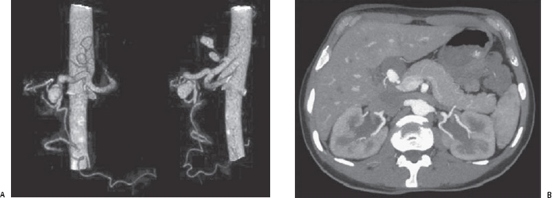
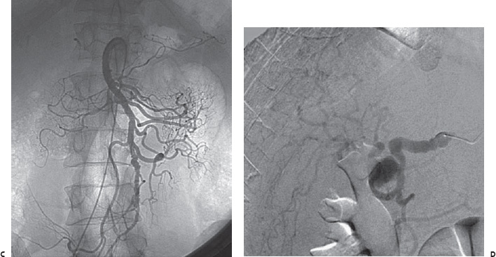
Case 70 An elderly man presents with abdominal pain. Further Work-up (A) Three-dimensional volume-rendered computed tomographic angiogram (CTA) shows saccular aneurysms of the hepatic (large arrow) and left gastric (small arrows) arteries. (B) Contrast-enhanced CT scan shows a thrombosed component of the hepatic artery aneurysm (arrows). (C) Selected superior mesenteric artery angiogram shows diffuse beading and focal segments of dilatation (arrows). (D) Selected hepatic angiogram shows beading of the common hepatic artery (arrow) and aneurysm of the proper hepatic artery (arrowhead). (E) Selected celiac angiogram shows successful coil embolization of the left gastric and hepatic aneurysms.

 Clinical Presentation

 Imaging Findings

Stay updated, free articles. Join our Telegram channel

Full access? Get Clinical Tree


