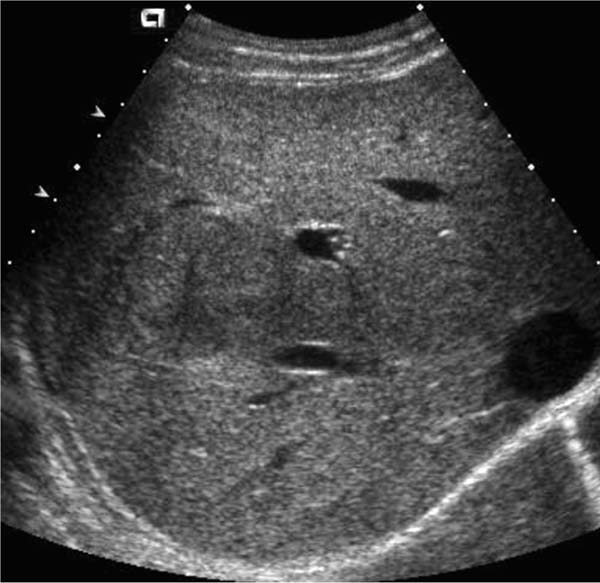
Case 70 A 35-year-old woman presents with vague right upper quadrant pain. Describe this follow-up study and amend the differential diagnosis if necessary. (A) Transverse ultrasound shows a well-circumscribed lesion (large arrow) that is slightly hypoechoic to liver with a hypoechoic center (small arrow). (B,C) T1 (B) and T2 fat saturation (C) axial magnetic resonance imaging (MRI): the lesion is slightly hypointense to liver on T1 with a hypointense center and hyperintense to liver on T2 (large arrows) with a hyperintense center (small arrows). • Focal nodular hyperplasia (FNH): In a young woman, a lesion that is slightly hypointense on T1 and slightly hyperintense on T2 with a very hyperintense central scar is most likely FNH. • Adenoma: This is second because of the high incidence and variable appearance. It may be hyperintense on T1, rather than hypointense like FNH, because of fat and glycogen. The vascular supply of adenoma is often draped at periphery, not in central scar as in FNH. • Fibrolamellar hepatoma: This is uncommon but may have a central scar and occur in young patients without cirrhosis.

 Clinical Presentation
Further Work-up

 Imaging Findings

 Differential Diagnosis
 Essential Facts
Stay updated, free articles. Join our Telegram channel

Full access? Get Clinical Tree


