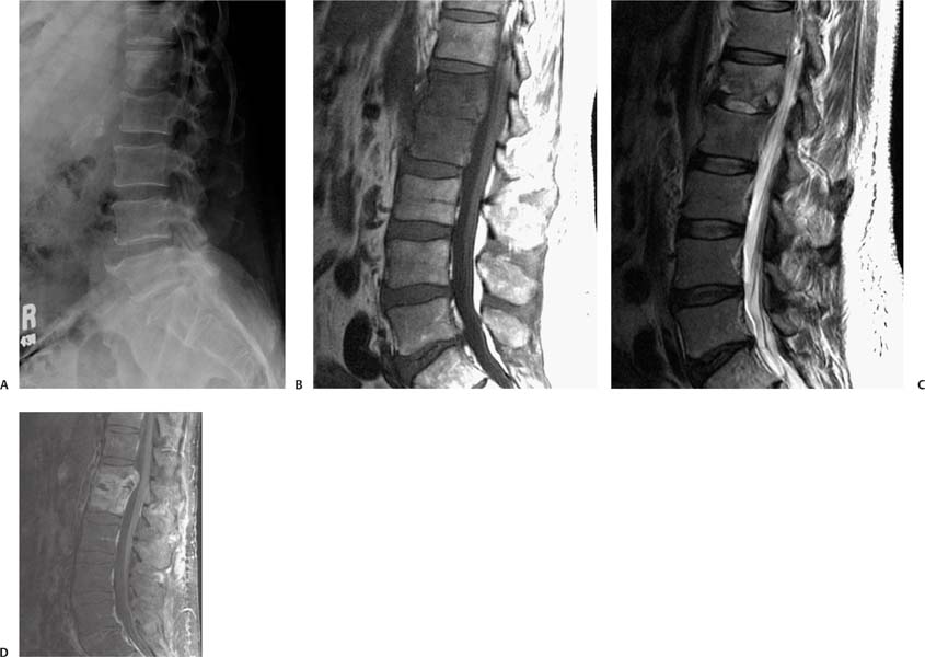
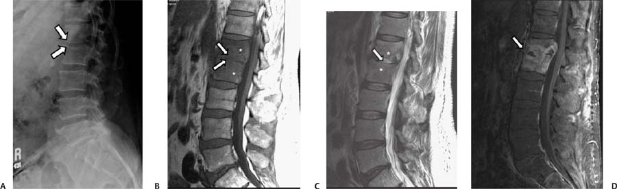
Case 71 A 59-year-old man with a 1-month history of worsening back pain. (A) Lateral view of the lumbar spine shows a decrease of the intervertebral space at L1-2 with poor definition of the adjacent end plates (arrows). (B) Sagittal T1-weighted image (WI) of the spine shows low signal in the L1 and L2 vertebral bodies (asterisks). The L1-2 disk shows low signal with poorly defined adjacent end plates (arrows). (C) Sagittal T2WI shows increased signal in the L1 and L2 vertebral bodies (asterisks) and in the L1-2 disk (arrows). (D) Sagittal fat-saturated T1WI of the lumbar spine with contrast shows diffuse enhancement of the L1 and L2 vertebral bodies and of the L1-2 disk (arrow). • Spondylodiskitis: This is pyogenic infection of the inter-vertebral disk and osteomyelitis of the adjacent vertebral bodies. The most frequent pathogen is Staphylococcus aureus
Clinical Presentation
Imaging Findings
Differential Diagnosis
![]()
Stay updated, free articles. Join our Telegram channel

Full access? Get Clinical Tree


