 Clinical Presentation
A 44-year-old woman with hemoptysis and hematuria.
Further Work-up

 Imaging Findings

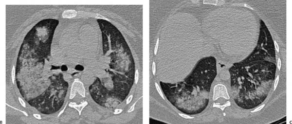
(A) Chest radiograph demonstrates patchy, lower lobe–predominant air-space opacity (black arrows). (B) Computed tomography (CT) of the chest (lung windows) at the level of the carina confirms multifocal ground-glass opacity (left arrow, posterior right arrow) with discrete nodules in the anterior right upper lobe and superior segment of the left lower lobe. The nodule in the right upper lobe has a ground-glass halo (anterior arrow). The nodule in the left lower lobe is cavitary. (C) CT of the chest (lung windows) at the lung bases (arrows) shows additional lower lobe consolidation and ground-glass opacity. There is no evidence of pleural or pericardial effusion, and the heart size is normal.
 Differential Diagnosis
• Wegener granulomatosis:
Stay updated, free articles. Join our Telegram channel

Full access? Get Clinical Tree


