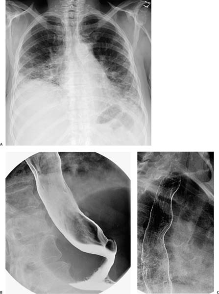
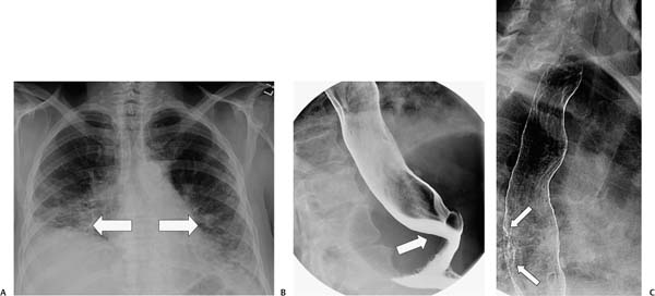
Case 71 A 68-year-old woman has gastroesophageal reflux and midepigastric pain. (A) Frontal chest radiograph shows bilateral basilar interstitial lung disease (arrows). (B) Upright barium esophagogram shows a widely patent gastroesophageal junction (arrow). (C) A focal segment of the dorsum of the esophagus shows mucosal irregularity (arrows), possibly representing esophagitis. • Scleroderma: This is the most likely diagnosis, given the patulous esophagus and the bilateral basilar interstitial lung disease. • Rheumatoid arthritis: This is a possibility as it can result in an atonic esophagus and interstitial lung disease.
 Clinical Presentation
 Imaging Findings
 Differential Diagnosis
Stay updated, free articles. Join our Telegram channel

Full access? Get Clinical Tree


