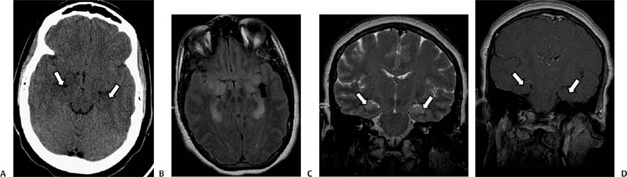
Case 72 A 37-year-old man with immunosuppression and acute neurologic deficit. (A) Axial computed tomography (CT) without contrast shows decreased density of the medial aspect of both temporal lobes (arrows). (B) Axial fluid-attenuated inversion recovery (FLAIR) image shows high signal in the medial aspect of both temporal lobes with a lack of gray–white matter differentiation (arrow). (C) Coronal T2-weighted image (WI) of the brain shows increased signal in the medial temporal lobes bilaterally (arrows). (D) Coronal T1WI of the brain with contrast shows patchy enhancement of the hippocampal region (arrows). • Herpes encephalitis:
Clinical Presentation
Further Work-up
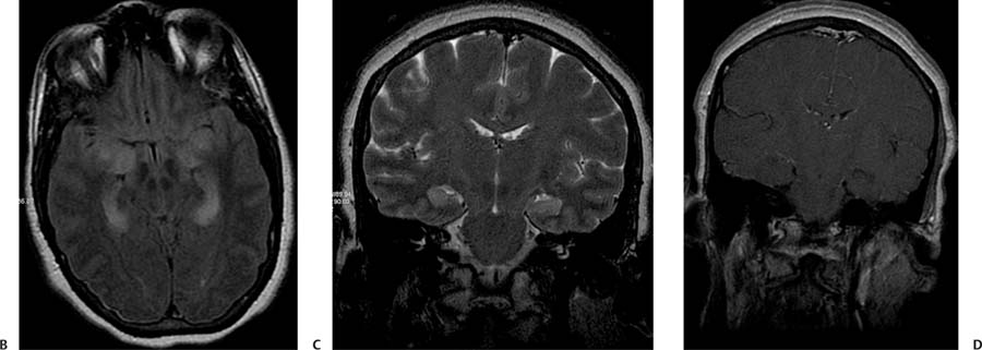
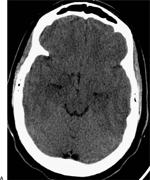
Imaging Findings
Differential Diagnosis
![]()
Stay updated, free articles. Join our Telegram channel

Full access? Get Clinical Tree


