 Clinical Presentation
A 50-year-old man with cough who failed treatment with antibiotics.
Further Work-up
 Imaging Findings
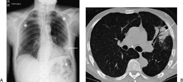
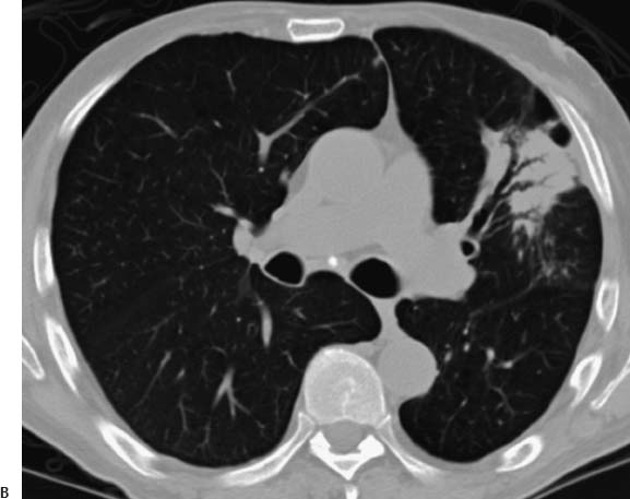
(A) Chest radiograph demonstrates peripheral left lung airspace opacity (arrow). (B) Computed tomography of the chest (lung windows) at the level of the carina (arrow) shows peripheral consolidation in the lingula.
 Differential Diagnosis
Stay updated, free articles. Join our Telegram channel

Full access? Get Clinical Tree


