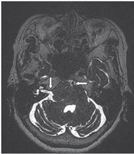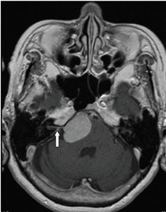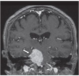


FINDINGS Figure 72-1. Axial T2WI through the cerebellopontine angle (CPA). There is a mass at the right CPA with a cleft of cerebrospinal fluid (CSF) separating it from the brainstem (arrow), confirming its extraaxial location. The adjacent brainstem is compressed and displaced to the left, demonstrating mild edema. The mass is ovoid, sharply circumscribed, and hypointense. Figure 72-2. Axial 3D volumetric heavily T2WI. This better demonstrates the relationship of the mass to the adjacent neural and vascular structures including cranial nerves VII and VIII (vertical arrow) and the basilar artery (BA) (transverse arrow). Figure 72-3. Axial post-contrast T1WI reveals a broad-based mass with intense, homogeneous enhancement with a dural tail extending into the right internal auditory canal (IAC) (arrow). Figure 72-4. Coronal post-contrast T1WI confirms the dural-based lesion along the tentorium cerebelli (arrow).
DIFFERENTIAL DIAGNOSIS
Stay updated, free articles. Join our Telegram channel

Full access? Get Clinical Tree


