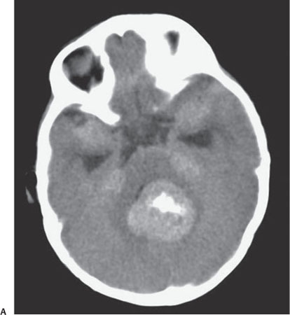
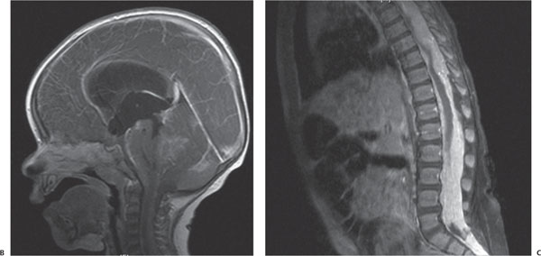
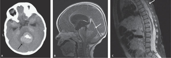
Case 72 A 3-year-old with ataxia. (A) Nonenhanced computed tomography (CT): there is a solid, hyperdense mass in the 4th ventricle with dense calcification (arrow). (B) Sagittal contrast-enhanced magnetic resonance image (MRI): there is an enhancing mass in the 4th ventricle (arrow) causing obstructive hydrocephalus. (C) Sagittal contrasted-enhanced MRI of the spine: there is extensive enhancing tumor within the spinal canal causing myelomalacia (arrow). • Medulloblastoma:
Clinical Presentation
Further Work-up
Imaging Findings
Differential Diagnosis
![]()
Stay updated, free articles. Join our Telegram channel

Full access? Get Clinical Tree


