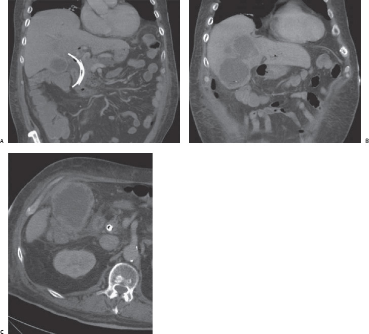
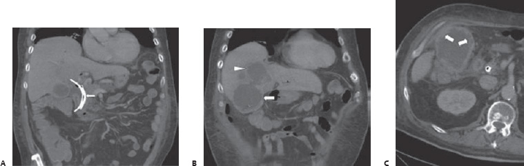
Case 72 A 79-year-old man who has pancreatic cancer presents with right upper quadrant pain 2 days after stent placement for malignant biliary obstruction. (A) Coronal contrast-enhanced computed tomographic (CT) scan shows metallic and plastic biliary stent s in place (arrow). (B) Thickening (arrow) and rupture of the gallbladder wall, resulting in a communicating hepatic abscess (arrowhead). The absence of gallstones indicates acalculous cholecystitis (AC). (C) Axial contrast-enhanced CT scan shows sloughing of the gallbladder epithelium (arrows), indicating gangrenous cholecystitis.
 Clinical Presentation
 Imaging Findings
 Differential Diagnosis
Stay updated, free articles. Join our Telegram channel

Full access? Get Clinical Tree


