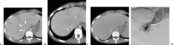
Case 72 A 23-year-old woman presents with abdominal distension. (A) Contrast-enhanced computed tomography (CT) shows heterogeneous peripheral enhancement (arrows), homogeneous central enhancement, and left lobe hypertrophy. (B) The right hepatic vein (arrow) appears occluded. (C) Delayed image shows homogeneous enhancement of the entire liver. (D) Transjugular hepatic venography shows the “spiderweb” pattern of hepatic veins, consistent with Budd-Chiari syndrome. • Budd-Chiari syndrome (BCS): This is the top choice for the diagnosis as it classically presents with delayed peripheral hepatic enhancement. This is further supported by the tortuous, narrowed right hepatic vein in Figure B. • Cirrhosis (without BCS): This can result in relative sparing of the left lobe, mottled enhancement, and tortuous vessels in the liver (“corkscrew” hepatic arteries). • Right heart failure:
 Clinical Presentation
 Imaging Findings
 Differential Diagnosis
![]()
Stay updated, free articles. Join our Telegram channel

Full access? Get Clinical Tree


