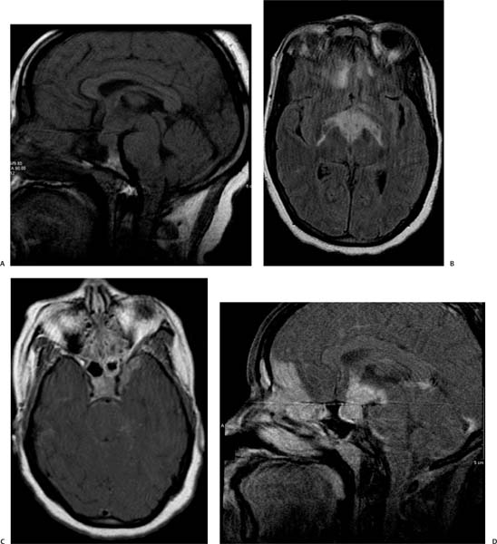
Case 73 A 37-year-old man with severe headache and palsy of the left abducens nerve. (A) Sagittal T1-weighted image (WI) shows a thickened pituitary stalk (white arrow) with effacement of the infundibulum (black arrow). (B) Axial fluid-attenuated inversion recovery (FLAIR) image shows high signal in the perimesencephalic cistern (arrows) and infundibular region (asterisk). There is also increased signal in the frontal lobes. (C) Axial T1WI with contrast shows enhancement in the sellar region (asterisk) that extends to the cavernous sinus on the left (arrow). (D) Sagittal T1WI with contrast shows enhancement that involves the sellar region (white arrow), infundibulum (black arrow
Clinical Presentation
Imaging Findings
![]()
Stay updated, free articles. Join our Telegram channel

Full access? Get Clinical Tree


