 Clinical Presentation
A 45-year-old woman who has mixed connective tissue disease presenting with cough and fatigue.
Further Work-up
 Imaging Findings
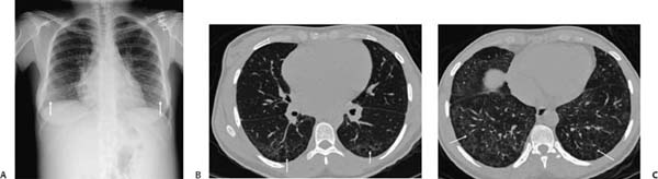
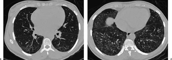
(A) Chest radiograph shows mild basilar opacity but is otherwise normal (arrows). (B) Computed tomography (CT) of the chest (lung windows) at the mid-thorax shows symmetric subpleural reticular opacities and micronodules (arrows). (C) CT of the chest (lung windows) at the lung bases shows more extensive reticular opacities, scattered areas of ground-glass opacity, and mild traction bronchiectasis (arrows).
Stay updated, free articles. Join our Telegram channel

Full access? Get Clinical Tree


