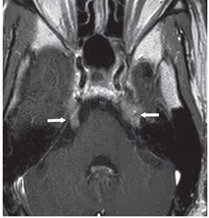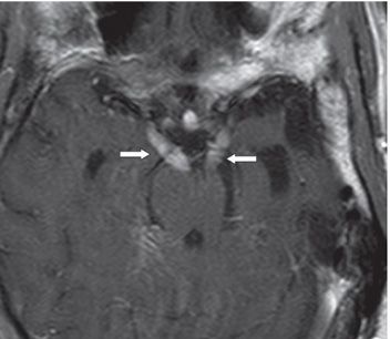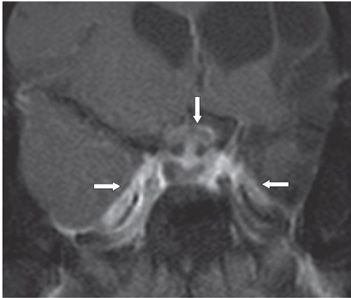


FINDINGS Figure 73-1. Axial FLAIR through the midbrain. There is thickening of the third cranial nerves bilaterally (arrows). Left brainstem atrophy is due to Wallerian degeneration from left middle cerebral artery (MCA) territory old infarct. There is thickening of the trigeminal nerves as well (not shown). Figure 73-2. Axial post-contrast T1WI through the brachium pontis. There is thickening and enhancement of the bilateral trigeminal nerves (arrows). Figure 73-3. Axial post-contrast T1WI through the midbrain. There is thickening and contrast enhancement of bilateral third cranial nerves (arrows). Figure 73-4. Coronal post-contrast T1WI through the sella. There is thickening and contrast enhancement of the chiasm and the infundibulum (vertical arrow). There is bilateral Meckel caves and cavernous sinuses enhancement (transverse arrows). There is a large left MCA territory encephalomalacia due to old cerebrovascular accident (line arrows).
DIFFERENTIAL DIAGNOSIS
Stay updated, free articles. Join our Telegram channel

Full access? Get Clinical Tree


