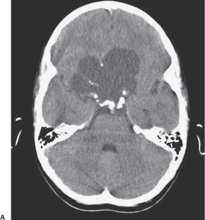
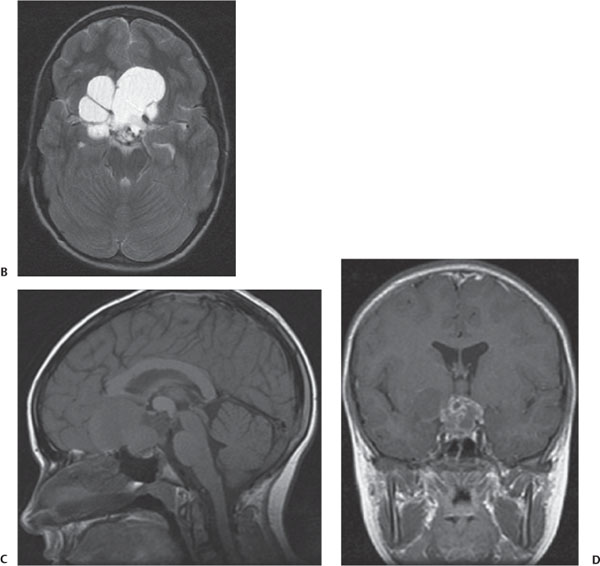
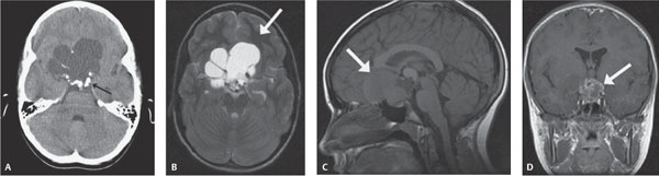
Case 73 A 10-year-old girl with headache and occasional vomiting. (A) Axial noncontrast computed tomography (CT) of the head: in the suprasellar region, there is a low-density, lobular mass with peripheral dense calcifications (arrow). (B) Axial T2-weighted magnetic resonance image (MRI): the lobular mass is fluid (arrow). (C) Sagittal T1-weighted MRI: the mass is intra- and suprasellar (arrow). (D) Coronal post-contrast MRI: there is peripheral enhancement (arrow). • Craniopharyngioma:
Clinical Presentation
Further Work-up
Imaging Findings
Differential Diagnosis
![]()
Stay updated, free articles. Join our Telegram channel

Full access? Get Clinical Tree


