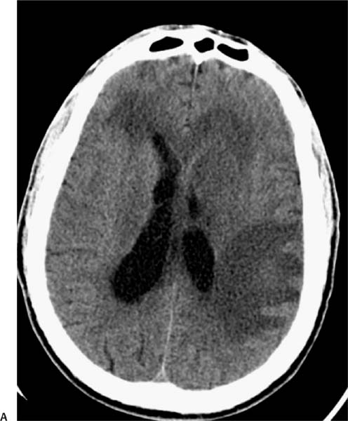
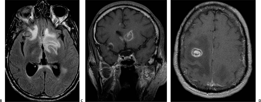
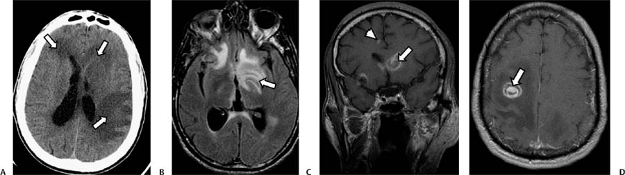
Case 74 A 42-year-old patient infected with human immunodeficiency virus presenting with deteriorating mental status. (A) Computed tomography (CT) of the brain without contrast shows areas of low attenuation in the white matter of both frontal lobes and in the left parietal region (arrows) related to vasogenic edema (the cortex is spared). There is effacement of the left frontal horn because of mass effect. (B) Axial fluid-attenuated inversion recovery sequence shows high signal in the frontal lobes bilaterally and in the left basal ganglia (arrow). (C) Coronal T1-weighted image (WI) with contrast shows a lesion with ring enhancement in the head of the left caudate nucleus (arrow) and a small enhancing lesion in the gray–white matter junction in the medial right frontal lobe (arrowhead). (D)
Clinical Presentation
Further Work-up
Imaging Findings
![]()
Stay updated, free articles. Join our Telegram channel

Full access? Get Clinical Tree


