 Clinical Presentation
A 62-year-old woman with progressive dyspnea.
Further Work-up
 Imaging Findings
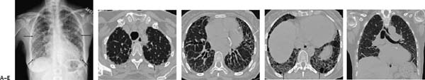
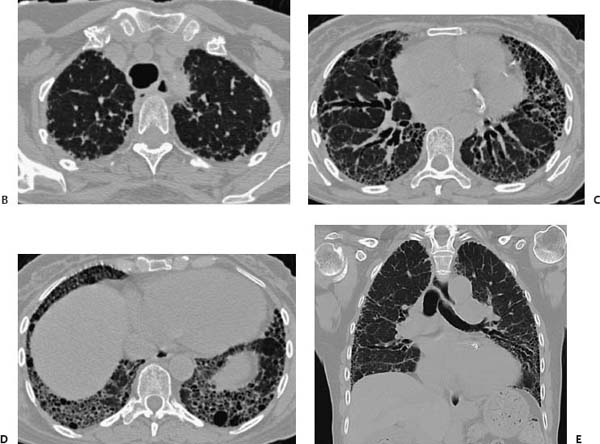
(A) Chest radiograph demonstrates symmetric base–predominant reticulation with volume loss (arrows). (B) Computed tomography (CT) of the chest (lung windows) through the lung apices demonstrates symmetric subpleural reticulation (arrow). (C) CT of the chest (lung windows) at the level of the aortic root shows more extensive traction bronchiectasis as well as honeycombing (arrows). There is no significant ground-glass component. (D) CT of the chest (lung windows) at the lung bases shows severe fibrosis and honeycombing (arrows). (E
Stay updated, free articles. Join our Telegram channel

Full access? Get Clinical Tree


