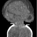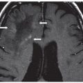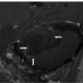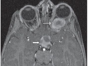
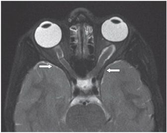
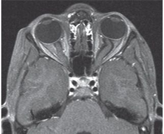
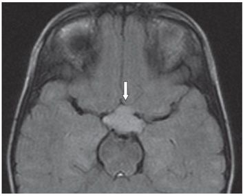
FINDINGS Figure 74-1. Axial T2WI through the orbits. There is expansion and hyperintensity of the left optic nerve with increased fluid under the dural sheath (arrow). The globe is proptotic and flat posteriorly. Figure 74-2. Axial post-contrast fat-suppressed T1WI through the orbits. There is enhancement of the optic nerve component (thin arrow) with a central hypointense nonenhancing portion. There is similar enhancing pattern in the chiasm (thick arrow). Figure 74-3. Axial T2WI in a companion patient. There is mild enlargement of both optic nerves (left greater than right) and no abnormal signal intensity. Figure 74-4. Axial post-contrast T1WI in the same patient. There is no contrast enhancement in either optic nerve. Figure 74-5. Axial FLAIR image through suprasellar cistern in the same patient. There is hyperintensity and lobulation of the optic chiasm (arrow).
DIFFERENTIAL DIAGNOSIS Optic neuritis, Optic pathway glioma (OPG) perioptic nerve schwannoma, optic nerve sheath meningioma, idiopathic orbital inflammatory pseudotumor, and sarcoidosis.
DIAGNOSIS Optic pathway glioma (OPG).
DISCUSSION
Stay updated, free articles. Join our Telegram channel

Full access? Get Clinical Tree


