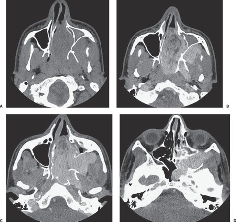
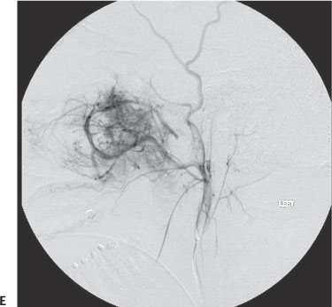
Case 74 A 15-year-old boy with nasal stuffiness. (A) Axial noncontrast computed tomography (CT): There is a soft-tissue mass filling the left nasal cavity and extending into the left nasopharynx (arrow). There is displacement of the posterior wall of the maxillary sinus and opacification of the maxillary sinus. (B–D) Axial contrast-enhanced CT images: there is dense contrast enhancement of the mass, which arises at the pterygopalatine fossa (PPF, arrows). (E) Anteroposterior digital subtraction external carotid arteriogram: there is a dense hypervascular tumor blush, with the major arterial supply from the distal left internal maxillary artery and its branches (arrow).
Clinical Presentation
Further Work-up
Imaging Findings
Differential Diagnosis
Stay updated, free articles. Join our Telegram channel

Full access? Get Clinical Tree


