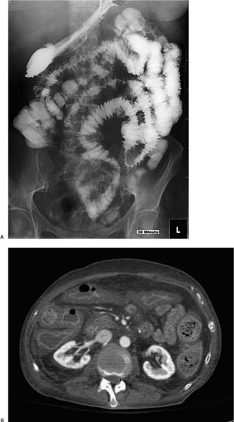
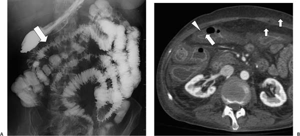
Case 74 A 52-year-old woman presents with abdominal pain. (A) Small-bowel follow-through shows diffuse, regularly thickened small-bowel folds (arrow). (B) Contrast-enhanced computed tomography (CT) in the arterial phase shows diffuse bowel wall thickening with a two-layer appearance: hyperdense mucosa (arrow) surrounded by a hypodense (water-density) submucosa (arrowhead). Small arrows indicate edematous fat. • Diffuse bowel wall edema:
 Clinical Presentation
 Imaging Findings
 Differential Diagnosis
![]()
Stay updated, free articles. Join our Telegram channel

Full access? Get Clinical Tree


