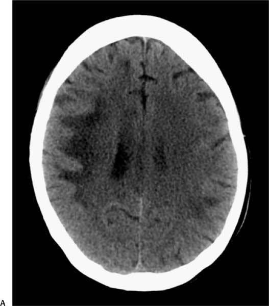
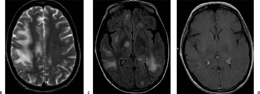
Case 75 A patient infected with human immunodeficiency virus presenting with headache, hemiparesis, and seizures. (A) Computed tomography (CT) of the brain without contrast shows asymmetric patchy areas of low attenuation bilaterally (arrows) with involvement of the subcortical U-fibers. (B) Axial T2-weighted image (WI) shows patchy scattered areas of increased intensity bilaterally, more prominent on the right side, involving the subcortical U-fibers (arrows). (C) Axial fluid-attenuated inversion recovery (FLAIR) image shows multiple patchy areas of hyperintensity involving the subcortical white matter and the right thalamus (arrow). (D) Magnetic resonance (MR) imaging of the brain with contrast shows no enhancement (arrow).
Clinical Presentation
Further Work-up
Imaging Findings
Differential Diagnosis
Stay updated, free articles. Join our Telegram channel

Full access? Get Clinical Tree


