 Clinical Presentation
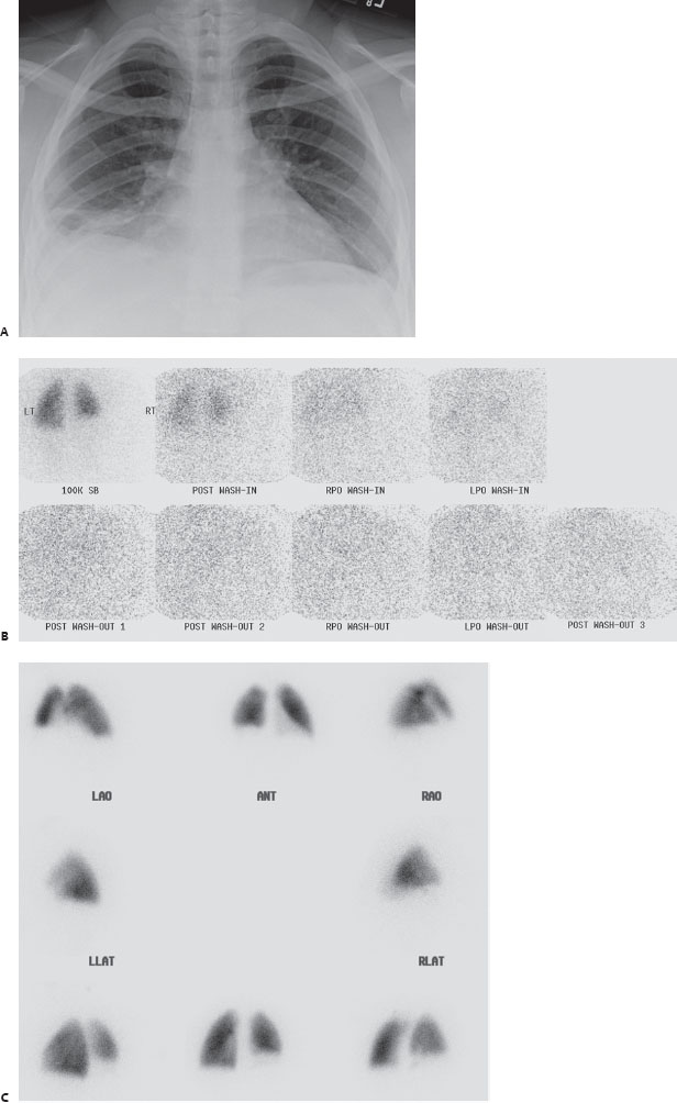
A 42-year-old woman with acute shortness of breath.

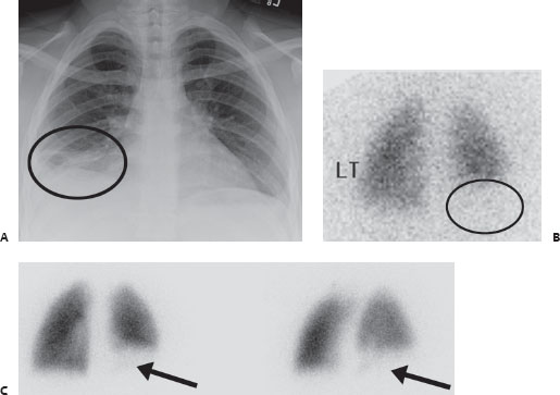
(A) Chest x-ray (CXR) demonstrates a small right pleural effusion and linear subsegmental atelectasis at the right lung base (circle). (B) Posterior and posterior oblique dynamic xenon 133 ventilation images demonstrate decreased ventilation to the right lung base on single breath (circle). No abnormal retention is seen on delayed imaging. (C) Multiple views from a Tc99m-MAA perfusion scan demonstrate a single large segmental perfusion defect in the posterior basal right lower lobe (arrows) that is matched to the ventilation and radiographic abnormalities (and is larger than the radiographic abnormality).
 Differential Diagnosis
• Intermediate probability for pulmonary embolism (PE) with evidence of pulmonary infarction:


Stay updated, free articles. Join our Telegram channel

Full access? Get Clinical Tree


