 Clinical Presentation
A 55-year-old man with cough. He is a heavy smoker.
Further Work-up
 Imaging Findings
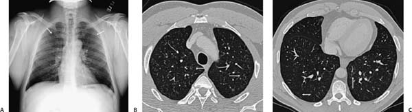
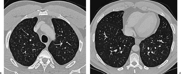
(A) Chest radiograph shows subtle, upper lobe–predominant, nodular interstitial abnormality (arrows). (B) Computed tomography (CT) of the chest (lung windows) at the lung apices demonstrates widespread bilateral centrilobular nodules on a background of centrilobular emphysema (arrows). (C) CT of the chest (lung windows) at the lung bases shows similar findings, but to a much lesser extent (arrows).
Stay updated, free articles. Join our Telegram channel

Full access? Get Clinical Tree


