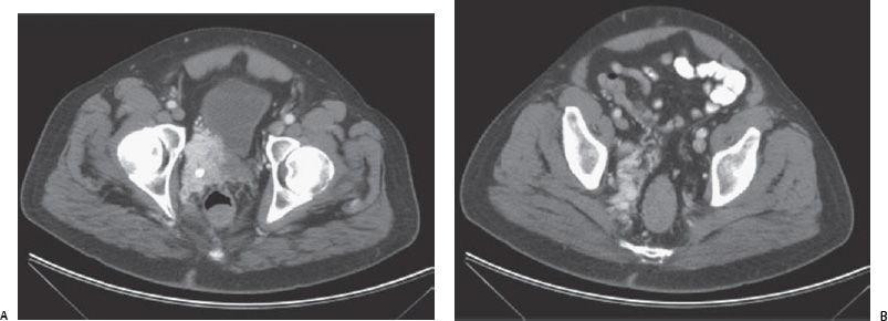
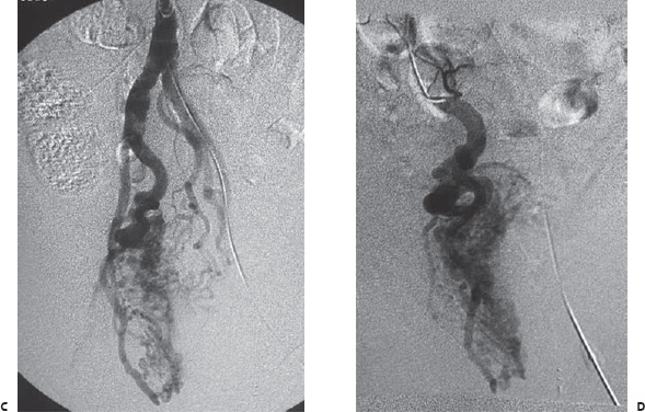
Case 75 A 37-year-old man presents to the clinic with a history of pelvic pain. (A) Infused computed tomographic (CT) scan shows enhancing mass abutting the bladder (arrows). The calcification is a phlebolith. (B) Higher cut shows right perirectal serpiginous structures (arrowhead). (C) Early phase pelvic angiogram shows the nidus (arrowhead) of a vascular malformation supplied by hypogastric (arrow) branches. (D) Selected hypogastric arteriogram shows large draining veins (arrow). (E) Multiple branches have been coiled and treated with N-butyl cyanoacrylate adhesive.

 Clinical Presentation
Further Work-up

 Imaging Findings

 Differential Diagnosis
Stay updated, free articles. Join our Telegram channel

Full access? Get Clinical Tree


