CASE 75
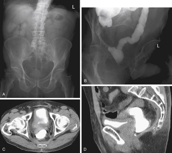
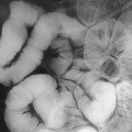
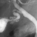
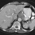
History: A 68-year-old man underwent ultra-low anterior resection 6 months ago. He had recovered well from his surgery. Earlier today he underwent a Gastrografin enema. He is now undergoing a CT scan without intravenous contrast.
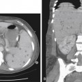
1. There is high-density material in the urinary bladder. Which of the following should be included in the differential diagnosis of this imaging finding as shown in Figures C and D? (Choose all that apply.)
C. Renal excretion of iodinated contrast administered intravascularly in the last few days
D. Vicarious renal excretion of iodinated contrast absorbed from the lumen of the intact colon
2. Several contributing factors that might increase the incidence of absorption of iodinated contrast from the colonic lumen have been recognized. Which of the following is not such a factor?
Stay updated, free articles. Join our Telegram channel

Full access? Get Clinical Tree


