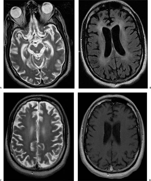
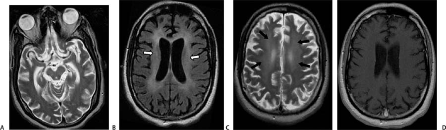
Case 76 A 40-year-old man infected with human immunodeficiency virus presenting with progressive dementia and tremor. (A) Axial T2-weighted image (WI) shows diffuse cerebral atrophy. (B) Axial fluid-attenuated inversion recovery (FLAIR) image shows diffuse cerebral atrophy with symmetric, confluent periventricular white matter disease (arrows). The subcortical white matter is spared. (C) Axial T2WI shows the symmetric periventricular white matter hyperintensities (arrows), with sparing of the subcortical U-fibers. (D) Axial T1WI with contrast shows no enhancement of the periventricular lesions. • Human immunodeficiency virus (HIV) encephalopathy:
Clinical Presentation
Imaging Findings
Differential Diagnosis
![]()
Stay updated, free articles. Join our Telegram channel

Full access? Get Clinical Tree


