 Clinical Presentation
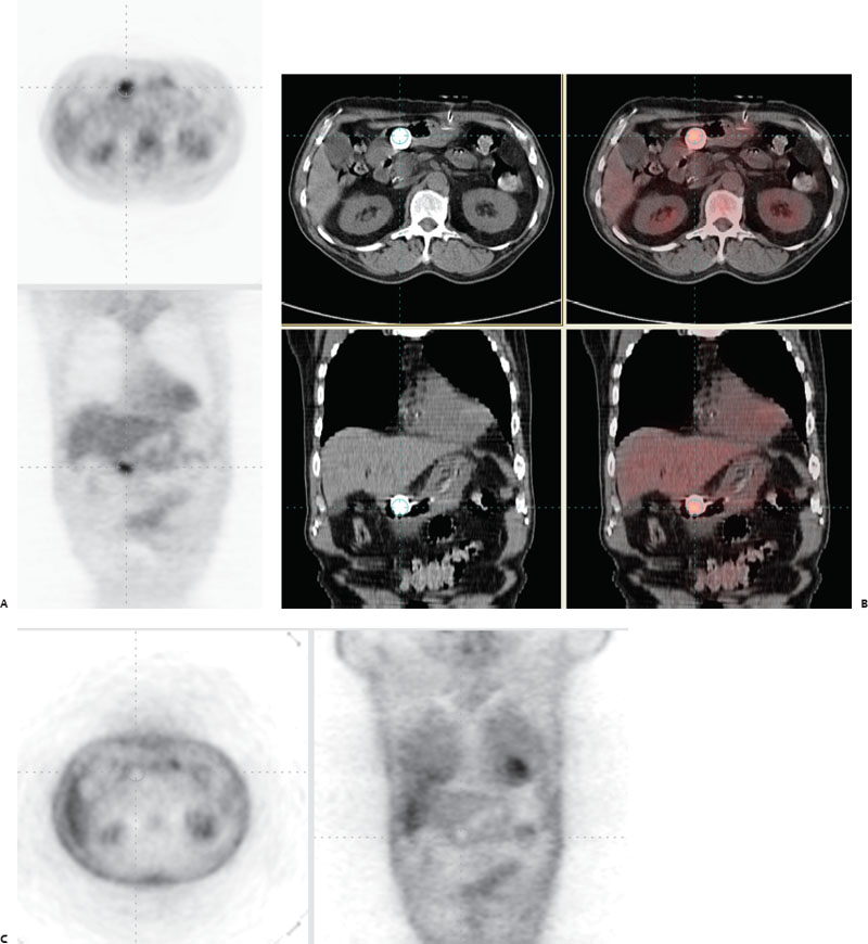
A patient presents for head and neck carcinoma restaging.

(A,B) Hybrid FDG-PET/CT imaging demonstrates a focal marked increase in radiotracer accumulation in the anterior abdomen (crosshairs). On CT, this corresponds to a contrast-filled gastrostomy catheter balloon. (C) PET emission image without attenuation correction demonstrates absent radiotracer accumulation in this same location.
 Differential Diagnosis
• Artifact of attenuation correction (AC): The lack of activity on the noncorrected images proves that this is an example artifactual “pseudo-uptake” of FDG.
• Metastasis to bowel: This can appear similar on AC images but will have a corresponding focus of some increased uptake on the noncorrected images.


Stay updated, free articles. Join our Telegram channel

Full access? Get Clinical Tree


