 Clinical Presentation
A 40-year-old woman with dyspnea and recurrent aspiration.
Further Work-up
 Imaging Findings
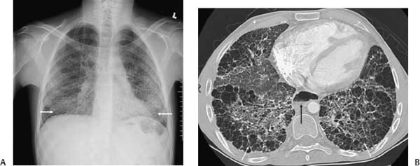
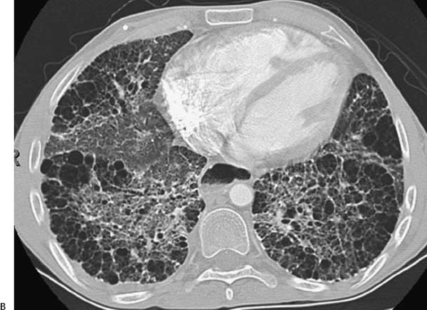
(A) Chest radiograph demonstrates lower lobe–predominant linear interstitial abnormality with small lung volumes (arrows). (B) Computed tomography of the chest (lung windows) at the lung bases shows extensive bilateral honeycombing and traction bronchiectasis (white arrows). The esophagus is severely dilated and contains an air-fluid level (black arrow
Stay updated, free articles. Join our Telegram channel

Full access? Get Clinical Tree


